
What we’ll cover
- What Is the Meniscus?
- Types of Meniscal Tears
- By Cause
- By Tear Pattern
- Causes and Risk Factors
- Signs and Symptoms
- How Is a Meniscal Injury Diagnosed?
- Clinical Examination
- Imaging
- Treatment for Meniscal Injuries
- Immediate First Aid
- Conservative (Non-Surgical) Treatment
- Surgical Treatment
- Recovery and Rehabilitation
- How to Prevent Meniscal Injuries
A meniscal tear is one of the most common knee injuries, affecting everyone from professional athletes to older adults doing everyday activities. Despite being so prevalent, many people are unsure what the meniscus actually is, what a tear feels like, and whether it always requires surgery. This guide covers everything you need to know.
What Is the Meniscus?
The knee is a hinge joint built for flexion, extension, and slight rotation. Inside it sit two C-shaped wedges of tough cartilage called the menisci, the medial (inner) and lateral (outer), positioned between the femur (thigh bone) and tibia (shin bone).
These structures do far more than simply cushion the joint. They absorb shock, distribute load evenly across the knee, improve joint stability, provide nutrition to the surrounding cartilage, and help control the range of movement. Without them, the joint surfaces would take a much greater pounding with every step.
The medial meniscus is more frequently injured than the lateral. Its shape and its firm attachment to the medial collateral ligament make it less mobile and more vulnerable to tearing under load.
Types of Meniscal Tears
Meniscal tears are classified in two ways: by what caused them and by the pattern of the tear itself.
By Cause
Traumatic tears are more common in younger, active people. They typically result from a twisting force applied to a weight-bearing, flexed knee and tend to follow a vertical or longitudinal pattern. The peak age of injury is 20 to 29 years, with men affected 2.5 to 4 times more often than women.
Degenerative tears occur in older adults as the meniscal tissue gradually weakens and loses its structural integrity over time. These are typically horizontal tears that can develop with minimal force, sometimes from something as unremarkable as an awkward twist when rising from a chair. Age-related changes also reduce the blood supply to the meniscus, which affects healing potential.
By Tear Pattern
Bucket-handle tear: a vertical or oblique tear that runs along the length of the meniscus, creating a loose flap that can displace into the joint and cause the knee to lock.
Radial tear: extends inward from the outer edge and disrupts the circumferential fibres responsible for load distribution.
Horizontal tear: a laminar split through the meniscal tissue; the typical pattern seen in degenerative injury.
Flap tear: a partial-thickness tear that creates a mobile fragment capable of catching between the joint surfaces.
It is also worth noting that sports-related meniscal injuries frequently occur alongside other knee injuries, particularly anterior cruciate ligament (ACL) tears.
Causes and Risk Factors
Meniscal tears result from a shear force between the femur and tibia. In younger patients, this is most commonly a twisting or pivoting movement on a planted foot with the knee slightly bent, such as during a sudden change of direction, a tackle, or a landing in sports like football, basketball, netball, or tennis. Deep or repeated squatting can also stress the meniscus.
In older patients, the cause is typically degenerative, driven by the progressive wear of joint cartilage over time. Habitual deep squatting or prolonged kneeling can accelerate this process.
Risk factors across all ages include participation in contact or pivoting sports, increasing age, obesity (which increases the compressive load on the joint), and a history of previous knee injury.
Signs and Symptoms
Symptoms vary considerably depending on the type and severity of the tear.
A traumatic injury may produce a popping or clicking sensation at the moment it occurs, followed by pain along the inner or outer joint line of the knee. Swelling typically develops over 24 to 48 hours. Stiffness, reduced range of motion, and difficulty fully straightening or bending the knee are also common.
Two distinct forms of locking can occur:
- Pseudo-locking is more common and refers to difficulty fully extending the knee due to swelling, pain, or muscle guarding rather than a physical obstruction.
- True locking is less common and occurs when a displaced bucket-handle fragment physically blocks full extension. The patient often has to perform a specific rotational movement to regain full range.
In smaller tears, initial symptoms may settle relatively quickly, and normal activity can resume, as the torn fragment does not always immediately interfere with joint mechanics. This can lead people to delay seeking treatment, which risks further damage.
Important: if symptoms are persistent or worsening, or if the knee will not extend fully, seek professional assessment promptly.
How Is a Meniscal Injury Diagnosed?
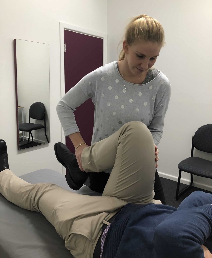
Clinical Examination
Diagnosis begins with a thorough history covering the mechanism of injury, when swelling appeared, any episodes of locking, activity level, and age. On examination, a physiotherapist or doctor will look for joint effusion (fluid), tenderness along the joint line, limited range of motion, and any quadriceps wasting in delayed presentations.
Several clinical tests are used to assess the meniscus:
- McMurray test: the knee is flexed then straightened with tibial rotation; pain, a click, or a clunk suggests a meniscal tear (sensitivity 10-66%, specificity 57-98%)
- Apley (grinding) test: performed prone with the knee at 90 degrees; axial compression combined with tibial rotation; pain or clicking suggests meniscal involvement
- Thessaly test: the most clinically sensitive option, with a sensitivity of 90% and specificity of 98%; the patient stands on the affected leg with the knee at 20 degrees of flexion and rotates the body; the test is positive if symptoms are reproduced
Imaging
An X-ray does not directly visualise the meniscus but is used to exclude fractures or osteoarthritis. MRI is the gold standard for diagnosing meniscal tears, providing detailed imaging of the soft tissue structures, including the menisci, cartilage, tendons, and ligaments, along with information about tear location and likely repairability.
CT arthrogram or ultrasound are alternatives when MRI is unavailable or contraindicated.
One important caveat: a tear seen on MRI does not automatically indicate that surgery is required. Clinical correlation with symptoms and function is always essential.
Treatment for Meniscal Injuries
Immediate First Aid
In the acute phase, follow the RICER protocol: Rest (offload the knee; crutches may be needed), Ice (20 minutes every two hours, wrapped in cloth, not applied directly to skin), Compression (elastic bandage to limit swelling), Elevation (leg raised above heart level), and Referral (to a physiotherapist or sports medicine professional as soon as possible).
Observe the No HARM guidelines for the first 48 to 72 hours: no heat, no alcohol, no running or activity, and no massage.
Conservative (Non-Surgical) Treatment
Conservative management is the appropriate first step for most patients, particularly those with no joint locking, minimal swelling, and a willingness to modify their activities. It is also the preferred approach for older adults with degenerative tears.
Activity modification is central: avoid twisting on a weight-bearing knee and temporarily cease aggravating sports. NSAIDs are recommended for 8 to 12 weeks to manage pain and inflammation; paracetamol is an alternative if NSAIDs are contraindicated.
Physiotherapy is the cornerstone of conservative management. A minimum of two sessions per week for at least eight weeks is recommended. The goals include normalising walking and range of motion, preventing quadriceps wasting, rebuilding strength and proprioception, and maintaining cardiovascular fitness during recovery.
The evidence for this approach is strong: studies of degenerative meniscal tears have demonstrated that supervised physiotherapy achieves equivalent pain reduction and functional improvement compared with partial meniscectomy combined with exercise.
Surgical Treatment
Surgery is reserved for patients with a locked knee, significant mechanical symptoms, or failure to improve with conservative management. It is also the preferred approach for younger patients with a vertical longitudinal tear in the outer, vascularised third of the meniscus, where surgical repair is most likely to succeed.
Procedures are performed via knee arthroscopy (keyhole surgery) and include:
- Meniscus repair: suturing of the torn pieces together; best outcomes in the well-vascularised outer zone; recovery takes 3 to 6 months
- Partial meniscectomy: removal of the damaged tissue only; allows early weight bearing with a shorter recovery of approximately 3 to 6 weeks; associated with a long-term increased risk of osteoarthritis
- Meniscal transplant: for younger active patients where significant meniscal tissue has been lost; uses donor tissue matched for size; performed at specialist centres only
A locked knee that will not extend is an absolute indication for surgical referral. Other indications include diagnostic uncertainty, failure to improve with conservative care, or persistent symptoms in any young patient.
Recovery and Rehabilitation
Recovery time varies significantly by tear type and treatment pathway. Minor tears managed conservatively typically improve over several weeks with adequate rest and physiotherapy. Partial meniscectomy carries a recovery of approximately 3 to 6 weeks before a return to activity, while meniscus repair requires 3 to 6 months due to the time needed for the torn tissue to re-unite.
Regardless of the pathway taken, rehabilitation aims to normalise gait, restore pain-free range of motion, prevent muscle wasting, rebuild strength and proprioception, and achieve a safe return to sport or pre-injury activities. With proper diagnosis, treatment, and a structured rehabilitation programme, most patients return to their pre-injury level of function.
How to Prevent Meniscal Injuries
While traumatic tears cannot always be avoided, several strategies meaningfully reduce risk:
- Train consistently before competition to ensure the body is adequately conditioned to handle the demands of sport
- Warm up properly before exercise and cool down afterwards
- Build strength, balance, coordination, and flexibility through a structured programme
- Increase training intensity and duration gradually rather than making sudden jumps in load
- Allow adequate recovery time between sessions
- Wear appropriate, supportive footwear for the activity to maintain knee stability
- Avoid overloading the knee in deep flexion, particularly with explosive or ballistic movements
- Modify activities that cause knee discomfort and be aware of the movements that aggravate the joint