When should I get a scan?
What we’ll cover
When should I get a scan?
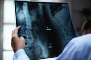
Medical technology is advancing rapidly year after year. Developments in medical imaging and scans (eg. MRI, CT, ultrasound etc.) provide clinicians with a huge amount of useful information regarding a patient’s presenting condition. As physiotherapists, in conjunction with a thorough assessment, we can use this information to guide our treatment plan and to judge if onward referral is needed. This ensures best practice within a multi-disciplinary setting.
Importantly, not every condition requires imaging to provide diagnosis or for complete recovery. In certain cases, imaging can be counterproductive and unnecessary. For example, we may be provided with irrelevant or excessive information on an image. Some of this information may not be relevant to a patient’s symptoms, pain or functional limitation.
In order to guide clinicians on interpretation of imaging reports and normal ageing, a number of recent studies have been done on pain free or asymptomatic individuals.
Can X-ray’s or MRI’s show issues in the shoulder, hip or back, even though the individual is pain free?
What does my shoulder ultrasound mean?
Ultrasounds are designed to produce images of soft tissue such as muscles, tendons and ligaments throughout the body. Ultrasounds are a common non-invasive imaging technique performed to look at the integrity and health of tendons and muscles around the shoulder.
In an ultrasound study of 50 middle aged men, 96% of them had “abnormal” findings on shoulder ultrasound, despite having no pain or disability. The most common abnormality detected was within a muscle of the rotator cuff known as the supraspinatus. 25% were shown to have a partial tear of supraspinatus yet were pain free and did not have any limitation. Some complete supraspinatus tears were also found within this group. These tears were detected only in individuals more than 60 years old.
The bursa was also identified on a number of these images as being “abnormal” on a number of these individuals. Commonly, the sub acromial, sub deltoid bursa is pinpointed as a source of pain in the shoulder. In the same study, 78% of participants had bursal thickening on ultrasound. Although the study was limited to a small group study of males only, it shows that as we approach middle age, bursal thickening and muscle tearing can be a normal finding on medical imaging [1].
Similar studies indicated a range of “abnormal findings” of the labrum in the shoulder. Recently, an increasing rate of surgical repairs have been performed on superior labral lesion repair. A 2016 study investigated the rate of superior labral lesions in the middle aged shoulder with no history of trauma. Over 55% of participants had superior labral tears on MRI! Importantly, none of these individuals had pain or disability [2]. However the majority of the population studied were sedentary.
What does an MRI show?
Magnetic Resonance Imaging (MRI) is a scanning procedure that uses magnets and radiofrequency to produce detailed pictures of soft tissue, bones and all internal body structures. MRI’s are commonly used for the diagnosis of hip and knee injuries.
A recent study utilised MRI to identify normal hip joint findings. Similar to the shoulder, there is also a labrum in our hip joint. The role of labral tissue is to act as a suction seal to hold our bone securely in the socket. The labrum also acts as a shock absorber, pressure distributer and joint lubricator. In a study of 40 Hip MRI’s of 15-66 year olds, 73% of individuals had some abnormality on their image. Labral defects were present in 69% of cases and cartilage defects in 24%. Of those found to have bony abnormalities (extra bone on the head of the femur), 95% of these individuals had labral defects. This extra bone is thought to pinch and damage our labral tissue as we move. It is often referred to as FAI (femoral acetabular impingement). However, how can this extra bone and labral damage be the only cause of pain and dysfunction in the hip, if it is commonly found in individuals who have no hip pain or disability? [3]
Even in the spine, degenerative changes are starting to be seen as signs of normal aging. In a review of 3110 spinal images (MRI, X-ray, CT scans), 90% of those aged over 60 years old had some abnormal finding on their scan. Issues such as disc degeneration, disc herniation and disc prolapse can start to become apparent, pain free, from the age of 20 years old. The 2014 review found that 30% of 20 year olds had signs of disc bulge, rising to 84% of 80 year olds. 31% of 30 year olds had evidence of disc protrusion on imaging, rising to 40% of those aged 70 years old. If these imaging findings do not relate to the level of a person’s symptoms, they should be regarded as normal aging findings.
When should I get a scan?
All of these statistics show high prevalence of abnormal imaging findings in people who are going about their daily lives completely unaffected by pain. Therefore, are there other issues we need to consider rather than just structure when it comes to musculoskeletal pain? Strength, flexibility, exercise tolerance, load management, pain beliefs may all play a role in how our bodies feel.
Another thought to ponder is: ‘are these pain free people with abnormal findings more likely to develop pain at a later stage?’ ‘Could we prevent this by optimising our body’s capacity to deal with the challenges of daily life?’
Your physiotherapist is well placed to perform a thorough assessment to identify likely causes and diagnosis of your musculoskeletal pain and symptoms. In addition, they can discuss your imaging results and the relationship to your symptoms.
Book online or call us today to see one of our physiotherapists who will be sure to point you in the right direction.
References
- Girish et al (2011), Ultrasound of the shoulder: asymptomatic findings in men, American Journal of Radiology (197)pp713-719
- Schwartzberg et al (2016), High Prevalence of Superior Labral Tears Diagnosed by MRI in Middle-Aged Patients With Asymptomatic Shoulders, The Orthopaedic Journal of Sports Medicine 4(1)
- Register et al (2012), Prevalence of Abnormal Hip Findings in Asymptomatic Participants: A Prospective, Blinded Study, American Journal of Sports Medicine (40)2720
- Brinjikjiet al (2014), Systematic Literature Review of Imaging Feature of Spinal Degeneration in Asymptomatic Populations, American Journal of Neuroradiology (10) 3174.


