Knee Conditions: Causes, Symptoms, Diagnosis and Treatment
What we’ll cover
- What Are Knee Conditions?
- Understanding the Knee Joint
- Key Structures of the Knee
- Common Causes of Knee Problems
- Traumatic Causes
- Overuse and Biomechanical Causes
- Degenerative and Medical Causes
- Symptoms of Knee Conditions
- Common Knee Symptoms and Symptoms by Location
- Warning Signs: When to Seek Urgent Care
- Common Knee Conditions and Injuries
- Patellofemoral Pain Syndrome (Runner’s Knee)
- Chondromalacia Patella
- Patellofemoral Maltracking
- ACL Injury (Anterior Cruciate Ligament Injury)
- Meniscus Tear
- MCL Tear (Medial Collateral Ligament Injury)
- Cartilage Damage and Microfracture Injuries
- Knee Osteoarthritis
- Patellar Tendinopathy (Jumper’s Knee)
- IT Band Syndrome
- Knee Bursitis
- Knee Fractures
- How Are Knee Conditions Diagnosed?
- Clinical Assessment
- Imaging and Investigations
- Knee Condition Treatment Options
- Conservative (Non-Surgical) Treatment
- Surgical Treatment Options
- Should I Use Ice or Heat on a Knee Injury?
- Knee Condition Recovery and Rehabilitation
- Recovery Timelines
- Key Principles of Knee Rehabilitation
- Exercises for Knee Conditions
- Exercises for Patellofemoral Pain Syndrome
- Exercises for Knee Osteoarthritis
- Exercises for Post-Ligament Injury and ACL Rehabilitation
- Stretches for Knee Pain
- Yoga for Knee Injuries
- Preventing Knee Conditions
- Lifestyle and Training Habits
- Biomechanical Risk Factors to Address
- Frequently Asked Questions
- What is patellofemoral chondromalacia?
- What are the most common knee conditions?
- Should I use ice or heat on a knee injury?
- How long does patellofemoral pain take to recover?
- What exercises help patellofemoral syndrome?
What Are Knee Conditions?
Knee pain is one of the most common musculoskeletal complaints in Australia, affecting people of all ages and activity levels. Whether you are an athlete, a tradesperson or someone managing the effects of ageing, knee problems can significantly impact your ability to move, work and stay active.
A knee condition refers to any disorder, injury or degenerative change affecting the structures of the knee joint. These conditions fall into three broad categories: traumatic injuries that occur suddenly (such as ligament tears or fractures), overuse conditions that develop gradually from repetitive loading (such as patellofemoral pain syndrome or patellar tendinopathy), and degenerative conditions driven by long-term wear and biological change (such as osteoarthritis).
Understanding the Knee Joint
Key Structures of the Knee
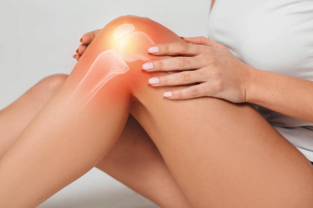
The knee is the largest and most complex joint in the human body. It connects the femur (thigh bone) to the tibia (shin bone), with the patella (kneecap) sitting at the front. Understanding the key structures helps explain why so many different conditions can develop and why the knee is particularly vulnerable to injury.
- Bones: the femur, tibia, fibula and patella form the framework of the knee joint
- Articular cartilage: smooth tissue covering the bone ends, allowing frictionless movement. It has a very limited capacity to self-heal.
- Menisci: the medial and lateral menisci are C-shaped fibrocartilage pads that act as shock absorbers between the femur and tibia
- Cruciate ligaments: the ACL and PCL run inside the knee and control forward, backward and rotational stability
- Collateral ligaments: the MCL (inner side) and LCL (outer side) resist sideways forces on the joint
- Patellofemoral joint: the joint between the kneecap and the front of the femur, responsible for guiding patellar movement during knee flexion and extension
- Tendons: the patellar tendon connects the patella to the tibia; the quadriceps tendon connects the quadriceps muscle to the patella
- Muscles: the quadriceps at the front and hamstrings at the back are the primary movers and stabilisers of the knee
- Bursae: small fluid-filled sacs that reduce friction between structures around the joint
Common Causes of Knee Problems
Traumatic Causes
Traumatic knee injuries occur suddenly from a specific event or mechanism, and are common in contact sports, falls and motor vehicle accidents.
- Sudden twisting, pivoting or change-of-direction movements under load
- Direct impact or tackle to the knee
- Falls, particularly onto hard surfaces
- Hyperextension of the knee joint
- Motor vehicle accidents
Overuse and Biomechanical Causes
Overuse injuries develop gradually when repetitive stress exceeds the tissue’s capacity to recover. Biomechanical factors can amplify this risk by placing an abnormal load on specific structures.
- Repetitive loading activities such as running, jumping and cycling
- Sudden increases in training volume or intensity
- Patellofemoral maltracking due to muscular imbalance or structural factors
- Weak hip abductors and gluteal muscles are causing dynamic valgus collapse
- Foot overpronation or high arches affecting lower limb alignment
- Inappropriate footwear for the activity or foot type
Degenerative and Medical Causes
Degenerative knee conditions are driven by long-term biological, mechanical and genetic processes, and are not solely the result of ageing.
- Age-related breakdown of articular cartilage
- Obesity increases joint loading forces
- Inflammatory conditions such as rheumatoid arthritis or gout
- Previous knee injuries that increase long-term osteoarthritis risk
- Genetic predisposition and family history of joint disease
- Occupations requiring prolonged kneeling, squatting or heavy lifting
Symptoms of Knee Conditions
Common Knee Symptoms and Symptoms by Location
Symptoms vary depending on which structures are affected and whether the onset was sudden or gradual. Identifying where in the knee you feel pain is one of the most useful early clues toward a diagnosis.
- Anterior (front) knee pain is commonly associated with patellofemoral pain syndrome, chondromalacia, patellar tendinopathy and patellar dislocation
- Medial (inner) knee pain is often linked to MCL tears, medial meniscus tears and medial compartment osteoarthritis
- Lateral (outer) knee pain is frequently caused by IT band syndrome, lateral meniscus tears or LCL injuries
- Posterior (back) knee pain may indicate a posterior horn meniscus tear, Baker’s cyst or PCL injury
General symptoms that can occur across many knee conditions include pain, swelling, stiffness (particularly after rest), clicking or grinding sensations, instability or a feeling the knee may give way, and reduced ability to fully bend or straighten the leg.
Warning Signs: When to Seek Urgent Care
Seek urgent medical attention if you experience any of the following:
- A loud pop or snap at the time of injury followed by immediate significant swelling
- Complete inability to bear weight on the affected leg
- Visible deformity of the knee joint
- A fever combined with a hot, red and swollen knee (possible infection)
- Numbness, tingling or loss of circulation below the knee
- A locking sensation where the knee physically cannot be straightened
More gradually, signs that your knees may need professional assessment include persistent morning stiffness, pain that worsens progressively over weeks, recurrent swelling after activity, and increasing difficulty with everyday tasks such as climbing stairs or rising from a chair.
Common Knee Conditions and Injuries
Patellofemoral Pain Syndrome (Runner’s Knee)
Patellofemoral Pain Syndrome (PFPS) is one of the most common knee conditions, accounting for up to 25% of all knee complaints presenting to physiotherapists. It refers to pain arising from the patellofemoral joint – the articulation between the kneecap and the front of the femur. Often called Runner’s Knee, it is particularly prevalent in runners, cyclists and young active adults, and is more common in females.
PFPS is characterised by a dull aching pain behind or around the kneecap, worsened by activities that load the patellofemoral joint such as stair climbing, squatting, kneeling and prolonged sitting. It is driven primarily by biomechanical and muscular factors and responds well to targeted physiotherapy and exercise rehabilitation.
Common causes: patellofemoral maltracking, weak hip abductors, tight quadriceps or IT band, sudden increases in training load, running on hard surfaces, foot overpronation.
Common symptoms: dull pain behind or around the kneecap, worsened by stairs and squatting, pain after prolonged sitting, occasional crepitus (grinding sensation), mild tenderness on palpation of the patellar borders.
Chondromalacia Patella
Chondromalacia patella refers to the softening, roughening and deterioration of the articular cartilage on the undersurface of the kneecap. It is a structural diagnosis, typically confirmed on MRI or arthroscopy, and is distinct from the symptom-based diagnosis of PFPS, though the two commonly co-exist. It is most common in young adults, particularly females, and in those with a history of patellofemoral maltracking or recurrent patellar dislocations. In older adults it may represent early patellofemoral arthrosis or patellofemoral arthropathy.
Common causes: patellofemoral maltracking, repetitive impact loading, history of patellar dislocation, direct trauma to the kneecap, age-related cartilage changes.
Common symptoms: pain behind or around the kneecap, grinding or grating sensation (crepitus), mild swelling, stiffness after rest, worsened by impact activities and stairs.
Patellofemoral Maltracking
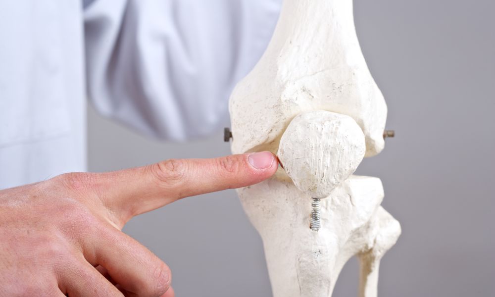
Patellofemoral maltracking describes the abnormal movement of the kneecap as it glides within the femoral groove during knee movement. Rather than tracking centrally, the patella typically deviates laterally, placing uneven stress on the cartilage and soft tissues of the patellofemoral compartment. It is a key contributing mechanism in PFPS, chondromalacia and patellar instability, and may be identified clinically via the J-sign during knee extension.
Common causes: weak VMO (inner quadriceps), tight lateral retinaculum or IT band, trochlear dysplasia, patella alta, weak hip abductors, foot pronation and lower limb malalignment.
Common symptoms: pain along the medial or lateral patellar border, patellar apprehension, pain with squatting or lunging, occasional partial dislocation (subluxation), J-sign on examination.
ACL Injury (Anterior Cruciate Ligament Injury)
An ACL injury is one of the most significant knee injuries in sport, particularly in activities involving sudden direction changes, pivoting and jumping. The anterior cruciate ligament runs diagonally inside the knee, controlling anterior tibial movement and rotational stability. Injuries range from mild sprains to complete ruptures, with complete tears most common in football, netball, basketball and skiing. The injury often occurs as a non-contact mechanism – a sudden deceleration or awkward landing – and is associated with the characteristic pop felt or heard at the time.
Common causes: sudden deceleration or pivoting, awkward landing from a jump, direct tackle forcing the knee inward, hyperextension, fatigue reducing neuromuscular control.
Common symptoms: audible or felt pop at the time of injury, immediate significant swelling (haemarthrosis), knee instability and giving way, inability to continue activity, restricted range of motion.
Meniscus Tear
The menisci are two C-shaped fibrocartilage pads sitting between the femur and tibia, functioning as shock absorbers and improving joint stability. Meniscus tears are among the most common knee injuries across both active and older populations. In younger patients they typically result from acute twisting injuries, often alongside an ACL injury. In older adults, degenerative tears can occur with minimal or no trauma. The medial meniscus is torn more frequently than the lateral.
Common causes: twisting or pivoting under load, deep squatting with rotational force, combined ACL injury, degenerative tearing in older adults, direct impact, prolonged occupational kneeling.
Common symptoms: pain at the medial or lateral joint line, swelling developing hours after injury, clicking, locking or catching sensations, difficulty fully bending or straightening the knee.
MCL Tear (Medial Collateral Ligament Injury)
The medial collateral ligament runs along the inner side of the knee and resists valgus forces that push the knee inward. MCL injuries are graded 1 to 3 based on severity and are among the most common ligament injuries in Australian contact sport. The most frequent mechanism is a direct blow to the outer knee. Unlike ACL injuries, isolated MCL tears generally have an excellent prognosis and often heal without surgery.
Common causes: direct blow to the outer knee, valgus stress during pivoting or landing, skiing falls, contact sport tackles, combined injury with ACL tear.
Common symptoms: pain and tenderness along the inner side of the knee, medial swelling and possible bruising, instability with sideways movement, pain on valgus stress testing.
Cartilage Damage and Microfracture Injuries
Articular cartilage covers the ends of the bones within the knee, enabling smooth movement. Because cartilage lacks a direct blood supply, it has a very limited capacity to self-repair. Damage can result from acute trauma, repetitive loading or degenerative disease. Full-thickness cartilage defects are a significant cause of long-term knee disability and are frequently identified on MRI, often alongside ACL injuries or meniscal tears.
Microfracture refers both to small stress fractures at the bone surface beneath damaged cartilage and to a surgical technique that creates perforations in the subchondral bone to stimulate fibrocartilage repair.
Common causes: acute joint trauma, patellar dislocation, repetitive high-impact loading, associated ACL injuries, age-related changes, inflammatory joint disease.
Common symptoms: deep joint pain often difficult to localise, swelling recurring with activity, clicking or catching within the joint, stiffness after inactivity, reduced load-bearing tolerance.
Knee Osteoarthritis
Knee osteoarthritis (OA) is the most common joint condition in adults over 45 and a leading cause of disability worldwide. It involves the progressive breakdown of articular cartilage, remodelling of subchondral bone, formation of bone spurs (osteophytes) and inflammation of the joint lining. OA can affect any compartment of the knee. Patellofemoral arthrosis and patellofemoral arthropathy specifically describe degenerative changes in the patellofemoral joint, presenting with anterior knee pain in middle-aged to older adults, and may be managed with physiotherapy, injections or patellofemoral joint replacement in appropriate cases.
Common causes: age, obesity, previous knee injury or surgery, occupational kneeling and heavy lifting, female sex particularly post-menopause, genetic predisposition.
Common symptoms: gradual onset joint pain with activity, morning stiffness lasting less than 30 minutes, bony joint enlargement, crepitus, reduced range of motion, possible varus or valgus deformity in advanced cases.
Patellar Tendinopathy (Jumper’s Knee)
Patellar tendinopathy is an overuse injury affecting the patellar tendon, which connects the kneecap to the tibia. It is particularly common in sports involving repetitive jumping and explosive loading such as basketball, volleyball, AFL and athletics. Pain is characteristically localised to the lower tip of the kneecap and is activity-related. Modern understanding recognises it as a degenerative rather than purely inflammatory process, which is why progressive loading rehabilitation is the cornerstone of treatment.
Common causes: repetitive jumping and landing, sudden training load increases, weak quadriceps and gluteals, hard playing surfaces.
Common symptoms: sharp pain at the inferior pole of the patella, pain during and after jumping or sprinting, localised tendon tenderness, stiffness with initial morning loading.
IT Band Syndrome
IT band syndrome (ITBS) is the most common cause of lateral knee pain in runners and cyclists. The iliotibial band is a thick connective tissue structure running from the hip down the outer thigh to the tibia. Repetitive knee movement can cause friction at the lateral femoral epicondyle, producing sharp or burning lateral knee pain. It is strongly associated with training volume errors and hip weakness, and rarely requires surgery.
Common causes: sudden increases in running volume, weak hip abductors and gluteals, excessive downhill running, tight lateral hip structures.
Common symptoms: sharp or burning pain on the outer knee, pain typically starting at a predictable point in a run, tenderness at the lateral femoral epicondyle, resolves with rest but returns on resumption of activity.
Knee Bursitis
Bursitis occurs when one of the small fluid-filled sacs (bursae) around the knee becomes inflamed, typically from repetitive pressure, direct trauma or prolonged kneeling. Common forms include prepatellar bursitis (in front of the kneecap, often called Housemaid’s Knee) and pes anserine bursitis on the inner side of the knee below the joint line.
Common causes: prolonged kneeling, direct impact to the kneecap, repetitive friction from sport, knee osteoarthritis, infection (septic bursitis).
Common symptoms: localised swelling and puffiness, tenderness on direct palpation, warmth and redness, pain with kneeling or direct pressure.
Knee Fractures
Fractures around the knee involve the patella, distal femur, proximal tibia (tibial plateau) or fibular head. Patellar fractures are the most common and typically result from a direct fall onto the knee. Tibial plateau fractures occur from high-energy axial loading. Stress fractures of the tibia can also develop in endurance athletes from repetitive loading rather than acute trauma. Most significant knee fractures require orthopaedic assessment and many require surgical fixation.
Common causes: direct fall onto the knee, motor vehicle accident, high-energy sporting collision, repetitive loading (stress fractures), osteoporosis in older adults.
Common symptoms: immediate severe pain, significant swelling, inability to weight-bear, possible visible deformity, bony tenderness on palpation.
How Are Knee Conditions Diagnosed?
Clinical Assessment
A thorough history and physical examination by a physiotherapist, sports physician or orthopaedic surgeon forms the foundation of diagnosis. This includes a detailed account of how and when the problem started, pain location and aggravating factors, and any relevant history of previous knee issues.
Physical assessment involves inspection for swelling and alignment, palpation of specific structures, range of motion testing and a series of validated special tests including the Lachman test (ACL), McMurray test (meniscus), valgus and varus stress tests (collateral ligaments), patellar apprehension test (patellofemoral instability) and Clarke’s sign (patellofemoral pain).
Imaging and Investigations
Imaging is used to confirm clinical findings and quantify the extent of injury. The most appropriate investigation depends on the suspected diagnosis.
- X-ray: shows bone structure, joint space narrowing, osteophytes and fractures. Cannot visualise soft tissue structures.
- MRI: the most comprehensive investigation for soft tissue injuries. Clearly shows ligaments, cartilage, menisci, bone marrow and tendons. Best used for ligament tears, meniscal tears and cartilage defects.
- Ultrasound: useful for tendon assessment, bursitis and guiding injections. Limited for deep joint structures.
- CT scan: provides detailed three-dimensional bone imaging. Used for complex fractures, trochlear dysplasia assessment and surgical planning.
- Bone scan: detects metabolic bone activity. Useful for stress fractures, infection and malignancy.
- Arthroscopy: direct visual inspection of the joint interior. Used for definitive diagnosis and simultaneous treatment where required, but as an invasive procedure is not used for diagnosis alone.
Knee Condition Treatment Options
Conservative (Non-Surgical) Treatment
The majority of knee conditions are initially managed conservatively, and many respond very well without the need for surgery.
- RICE / PEACE and LOVE protocol: first-line management for acute injuries to reduce pain and swelling. Modern guidance favours Protection, Elevation, Avoiding early anti-inflammatories, Compression and Education, followed by progressive Load, Optimism, Vascularisation and Exercise.
- Physiotherapy and rehabilitation: the cornerstone of most knee condition management, including exercise prescription, manual therapy, neuromuscular retraining and structured return-to-sport programs.
- Patellofemoral taping (McConnell taping): a specific taping technique to correct patellar maltracking and reduce anterior knee pain. Evidence supports short-term pain reduction when combined with exercise for PFPS and chondromalacia.
- Bracing and orthotics: patellar tracking braces for patellofemoral conditions; hinged braces for ligament injuries; foot orthotics to correct overpronation contributing to lower limb malalignment.
- Anti-inflammatory medications: oral or topical NSAIDs to reduce pain and swelling in acute inflammatory presentations. Short-term use under medical guidance.
- Corticosteroid injections: anti-inflammatory injections providing short-to-medium term pain relief for OA, bursitis and inflammatory conditions.
- Hyaluronic acid injections: viscosupplementation injections used in knee OA, most effective for mild-to-moderate disease alongside exercise therapy.
- PRP injections: platelet-rich plasma injections using the patient’s own growth factors to support healing in tendinopathy and OA.
- Activity modification and load management: structured reduction of aggravating activities to allow tissue recovery, particularly important for overuse conditions.
- Weight management: reducing body weight significantly reduces forces through the knee joint and is one of the most effective interventions for knee OA.
Surgical Treatment Options
Surgery is reserved for structural injuries that fail conservative management, significant instability, or end-stage degenerative disease.
- ACL reconstruction: rebuilding the torn ACL using a graft (typically hamstring or patellar tendon). Recommended for active patients seeking return to sport and those with ongoing instability.
- Meniscal repair or meniscectomy: arthroscopic repair of torn meniscal tissue where possible, or partial removal of irreparable tears.
- Microfracture surgery: creating small perforations in subchondral bone to stimulate fibrocartilage repair at full-thickness cartilage defects.
- Osteochondral autograft/allograft (OATS / OCA): transplantation of cartilage and bone to resurface significant osteochondral defects; provides superior outcomes to microfracture for larger lesions.
- MPFL reconstruction: reconstruction of the medial patellofemoral ligament for recurrent patellar dislocation and instability.
- Tibial Tubercle Osteotomy (TTO): repositioning of the tibial tubercle attachment to correct patellofemoral maltracking and reduce joint stress.
- Partial knee replacement: resurfacing of a single knee compartment; preserves more native joint tissue than total replacement.
- Total knee replacement (TKR): complete resurfacing of the knee joint; gold-standard treatment for end-stage symptomatic knee OA.
- Patellofemoral joint replacement: replacement of the patellofemoral compartment only, appropriate for isolated patellofemoral arthrosis or arthropathy.
Should I Use Ice or Heat on a Knee Injury?
Use ice during the first 48 to 72 hours after an acute injury or during a flare-up of active swelling and inflammation. Apply for 15 to 20 minutes at a time with a cloth barrier to protect the skin.
Use heat for chronic stiffness or muscular tightness, particularly before activity or physiotherapy sessions. Heat is appropriate for long-standing conditions such as osteoarthritis when there is no active swelling present.
Avoid heat on a freshly injured or actively swollen knee, as it can increase inflammation. If you are unsure which is appropriate for your condition, consult your treating physiotherapist or clinician.
Knee Condition Recovery and Rehabilitation
Recovery Timelines
Recovery timelines vary considerably depending on the condition, severity and treatment pathway. The following provides general guidance only.
- Patellofemoral Pain Syndrome: 6 to 12 weeks with physiotherapy; return to sport 6 to 12 weeks
- ACL Tear (complete): 9 to 12 months post-reconstruction; return to sport 9 to 12 months
- Meniscus Tear (repair): 4 to 6 months post-surgery; return to sport 4 to 6 months
- Meniscus Tear (partial meniscectomy): 6 to 12 weeks post-surgery
- MCL Tear Grade 1 to 2: 4 to 12 weeks of conservative management
- MCL Tear Grade 3: 3 to 6 months conservative or surgical
- Patellar Tendinopathy: 3 to 6 or more months with progressive loading rehabilitation
- Total Knee Replacement: 6 to 12 weeks to return to daily activities
Key Principles of Knee Rehabilitation
- Progressive loading: gradually increasing stress through the healing tissue guides recovery without overloading
- Pain-guided activity: Some discomfort during exercise is acceptable; sharp or persisting pain after exercise signals overloading
- Quadriceps and gluteal strengthening: essential for almost all knee conditions, improving joint stability and reducing abnormal loading
- Neuromuscular control and proprioception: retraining joint position sense and movement patterns significantly reduces re-injury risk
- Criteria-based return to sport: Objective tests such as hop tests and strength symmetry assessments should guide return to sport, not time alone
- Psychological readiness: fear of re-injury is as important as physical readiness, particularly following ACL reconstruction
Exercises for Knee Conditions
Exercise therapy is central to the management of almost all knee conditions. The following exercises are commonly prescribed by physiotherapists. Always consult a qualified physiotherapist before commencing a rehabilitation program, as exercise selection must be tailored to your specific diagnosis and stage of recovery.
Exercises for Patellofemoral Pain Syndrome
These exercises target the muscular deficits most commonly associated with patellofemoral conditions, including weak VMO, weak hip abductors and poor lower limb neuromuscular control.
- Terminal Knee Extension (TKE): activates the VMO near full extension with low patellofemoral joint stress; foundational exercise for PFPS
- Short Arc Quads: quadriceps activation through a limited pain-free arc; suitable for early rehabilitation
- Straight Leg Raise (SLR): strengthens the quadriceps without bending the knee; minimal patellofemoral loading
- Step-Downs (eccentric): eccentric quadriceps and gluteal loading; trains movement control and addresses kinematic deficits
- Clamshells and Hip Abduction: strengthens gluteus medius; reduces valgus collapse and abnormal patellar tracking
- Single Leg Press (partial range): progressive lower limb loading; range adjusted to remain pain-free
- Wall Slides (shallow squats): controlled patellofemoral loading; depth adjusted to the patient’s pain-free range
Exercises for Knee Osteoarthritis
- Low-impact strengthening: leg press, stationary cycling and hydrotherapy
- Quadriceps and gluteal exercises to support and offload the joint
- Avoiding high-impact activities such as running on hard surfaces during symptomatic flare-ups
Exercises for Post-Ligament Injury and ACL Rehabilitation
- Early phase: restore range of motion, manage swelling, activate quadriceps
- Mid phase: progressive lower limb strengthening, proprioception and balance training
- Late phase: sport-specific movement retraining, plyometric loading and running progressions
- Return-to-sport testing: single-leg hop tests and quad and hamstring strength symmetry assessments
Stretches for Knee Pain
Hold each stretch for 30 to 45 seconds and repeat 2 to 3 times per side.
- Quadriceps stretch: standing or prone, heel drawn toward the gluteal
- Hamstring stretch: seated or supine with gentle knee extension
- Hip flexor stretch: low lunge position with the hip gently pressed forward
- IT band stretch: standing cross-leg stretch or foam roller along the outer thigh
- Calf stretch: wall stretch with both straight and bent knees to target the gastrocnemius and soleus
Yoga for Knee Injuries
Yoga can complement a knee rehabilitation program by improving flexibility and body awareness. Poses generally well-tolerated with most knee conditions include low lunge, supported bridge, reclined pigeon and modified warrior I. Avoid deep knee flexion poses such as full lotus or hero pose during an active knee condition, and always inform your yoga teacher about your injury before class.
Preventing Knee Conditions
Lifestyle and Training Habits
- Maintain a healthy body weight to minimise joint loading
- Build and maintain strong quadriceps, gluteal and hamstring muscles through regular progressive strength training
- Avoid sudden large increases in training volume or intensity; follow the 10% weekly increase rule as a guide
- Warm up adequately before sport and allow time for a structured cool-down and stretch afterwards
- Use appropriate and well-fitting footwear for your activity and foot type
- Allow adequate recovery between sessions and avoid chronic overtraining
- Incorporate evidence-based injury prevention programs such as the FIFA 11+ warm-up (proven to reduce knee injuries in football), the Netball Australia KNEE program and ACL prevention protocols used by Australian football codes
Biomechanical Risk Factors to Address
- Foot pronation and inappropriate footwear contribute to lower limb malalignment
- Hip abductor weakness allows the knee to collapse inward during loading
- Genu valgum (knock knees), sometimes referred to as knee knock disease, is a structural condition where the knees angle inward. In children, a degree of valgus is developmentally normal and typically self-corrects; in adults, persistent valgus increases patellofemoral stress and lateral knee loading and warrants physiotherapy assessment
- Structural patellar abnormalities, such as patella alta or trochlear dysplasia, predisposing to maltracking
Frequently Asked Questions
What is patellofemoral chondromalacia?
Patellofemoral chondromalacia, also known as chondromalacia patella, refers to the softening and deterioration of the cartilage on the undersurface of the kneecap. It is a structural finding confirmed on MRI or arthroscopy, graded I to IV based on severity. It is related to but distinct from patellofemoral pain syndrome, which is a symptom-based diagnosis. The two conditions commonly co-exist.
What are the most common knee conditions?
The most common knee conditions include knee osteoarthritis (most prevalent in adults over 45), patellofemoral pain syndrome (most common in active young adults), meniscus tears, ACL injuries, patellar tendinopathy and IT band syndrome. MCL tears are among the most common ligament injuries in contact sports.
Should I use ice or heat on a knee injury?
Use ice for acute injuries and active swelling during the first 48 to 72 hours. Use heat for chronic stiffness and muscular tightness when no active inflammation is present. Avoid heat on a freshly injured or swollen knee. When in doubt, consult your physiotherapist.
How long does patellofemoral pain take to recover?
Most people experience significant improvement within 6 to 12 weeks of a structured physiotherapy program. Long-standing PFPS or associated chondromalacia may require several months of management, with some individuals requiring ongoing load monitoring to prevent recurrence.
What exercises help patellofemoral syndrome?
The most evidence-supported exercises target hip abductor strengthening (clamshells, hip abduction), VMO activation (terminal knee extension, short arc quads) and lower limb movement control (step-downs, single leg press). Quadriceps, IT band and hip flexor stretching is also recommended. Exercises should be prescribed specifically by a physiotherapist based on your individual capacity and biomechanical assessment.